
Typically, a routine EEG is performed on an alert adult patient with recording time of 20 minutes plus 3 minutes hyperventilation and photic stimulation. There are many instances when deviating from the routine procedure is required.
Clinical reasons to alter from the routine procedure include when the patient may be drowsy, unconscious or have altered consciousness. In these instances, we may need to adapt the routine procedure by extending the recording time to suit the needs of the patient and gain the most information from the recording. Electrographically there may be reasons to change the routine procedure; if a particular pattern is difficult to localise it may be helpful to add additional electrodes.
Even in the alert patient there may be reasons to vary the EEG from the routine procedure. It is important to interpret the patient’s needs, request information and the EEG recording individually. The technologist/scientists ability to vary the procedure will allow the most information available to the reporting physician.
Changes to the routine procedure according to age
The EEG is adapted to the needs of the patient. Different techniques and strategies are used to perform an EEG on a baby than those used for a toddler or a young child. Although there are specific procedures necessary to record a comprehensive EEG it is important to ‘follow the lead’ of the patient to a certain degree to obtain the best possible recording. A young child may refuse to close their eyes. A readable recording with minimal eye closure is much better than an EEG recording on a distressed patient refusing to co-operate.
Additional recording electrodes
There may be times when additional electrodes applied will help clarify the focus or the source of the waveforms.
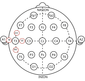
Additional electrodes can be added when an equipotential discharge is suspected. This electrode placement diagram shows additional electrode positions according to the 10/10 electrode placement system, an adaption to the 10/20 electrode placement system.
In picture below there are three snapshots of the same EEG epoch with spike and wave discharges appearing in the left central and temporal region.
The first snapshot of EEG in an Anterior – Posterior montage shows phase reversing spike and wave discharges at C3 and T3.
In order to define the focus a little better, the transverse montage is chosen. In this second snapshot the spike and wave discharges now phase reversal across the top of C3-T3 but no clear spike and wave activity at either C3 or T3.
By adding additional EEG recording electrodes between C3 and T3 (electrode 25) and T3 and A1 (electrode 31) as seen in the third snap shot, a clear phase reversal is seen at electrode 25.
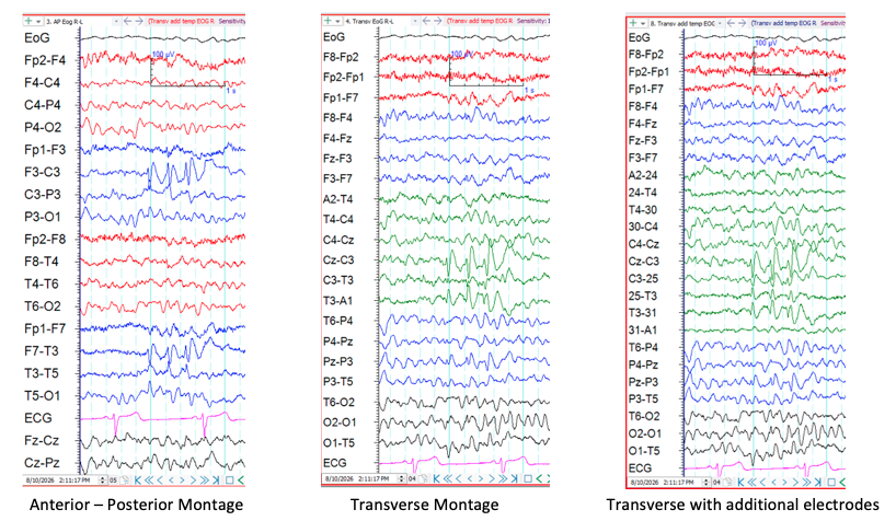
Reminder: Differential amplifiers only amplify the difference between the two inputs so any signal presenting equally will not be amplified. In this case the spike and wave activity were equally represented at C3 and T3 so were no amplified at the C3-T3 EEG recording channel.
You may wish to revisit the ANSA Learning short course Neurophysiology Amplifiers, Filters and Output
EEG recording derivations are discussed in more detail in the ANSA Learning short course EEG Recording derivations(as at Sept 2026 availability TBA).
Changes to Activation Procedures
There will be instances when changing the activation procedure can be helpful. Hyperventilation is known to most likely induce an absence seizure on a young child who has Juvenile Absence Epilepsy. If an absence seizure has not been induced within the usual 3 minute protocol of hyperventilation, extending to 5 minutes may increase the yield.
Patient’s with a photo-paroxysmal response should be re-presented with the responsive flash frequencies to identify the reproducibility and define the offending flash frequencies. Monocular stimulation or introducing polaroid glasses to see if the paroxysmal activity attenuates can also be added to the procedure and can assist with treatment plans.
Sleep deprivation is another activation procedure that can increase the yield of the recording not only by putting the brain under stress due to lack of sleep but also increasing the chance of the patient being drowsy and drifting into light sleep. Sometimes patients may not be sleep deprived but are a little drowsy. By recording for say, 10 minutes longer than usual, you may capture the patient drifting off to sleep. The drowsy period is a vulnerable time for the brain and is often when epileptiform activity appears. Recording for 20 minutes should not be the hard and fast rule, simply the minimum recording time.
A drowsy patient may need to be woken to the alert state for part of the recording. Simple techniques like asking the patient to count out loud to see if there is measurable posterior dominant rhythm.
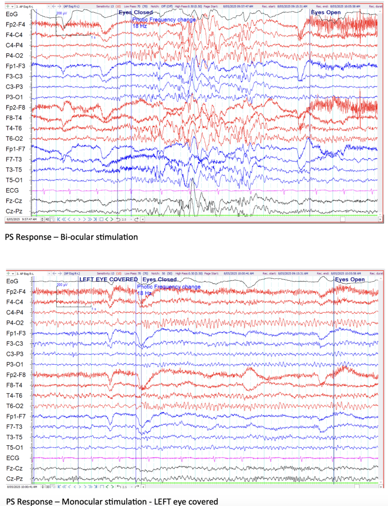
In the example above, the same patient is presented with the photic stimulation at 18Hz on two separate occasions. The first sample shows a burst of generalised spike and wave activity with both eyes closed. The second sample shows no photo-paroxysmal response with the both eyes closed and the left eye covered.
