
The electrocardiogram (ECG) should be recorded routinely with the EEG. This allows correlation of ECG and pulse rhythms to the EEG as well as assisting interpretation of the EEG when cardiac symptoms are involved.
Other physiological measures in the routine EEG recording may include respiration, EMG and eye movement. Other physiological measures are discussed in more detail in the ANSA Learning short course Additional Physiological Recording Techniques (as at Sept 2026 availability TBA).
ECG – Electrocardiograph
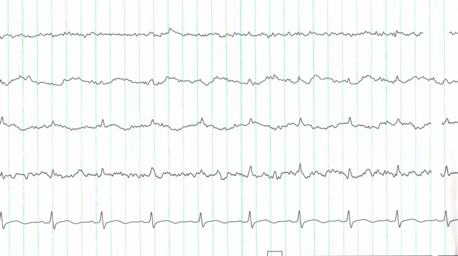
The recording above is a good example of when the ECG trace can help differentiate pulse and ECG artefact from the background EEG.
EOG – Eye movement monitoring – Electro-oculogram
The technologist/scientist may come across particular activity that is difficult to differentiate between eye movement and frontal cerebral activity. EOG electrodes can be useful to determine the difference
A simple one channel EOG with electrodes placed at the outer canthus of each eye, one slightly above and one slightly below can provide a good representation of both horizontal and vertical eye movement. EOG = R eye – Left Eye.
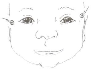
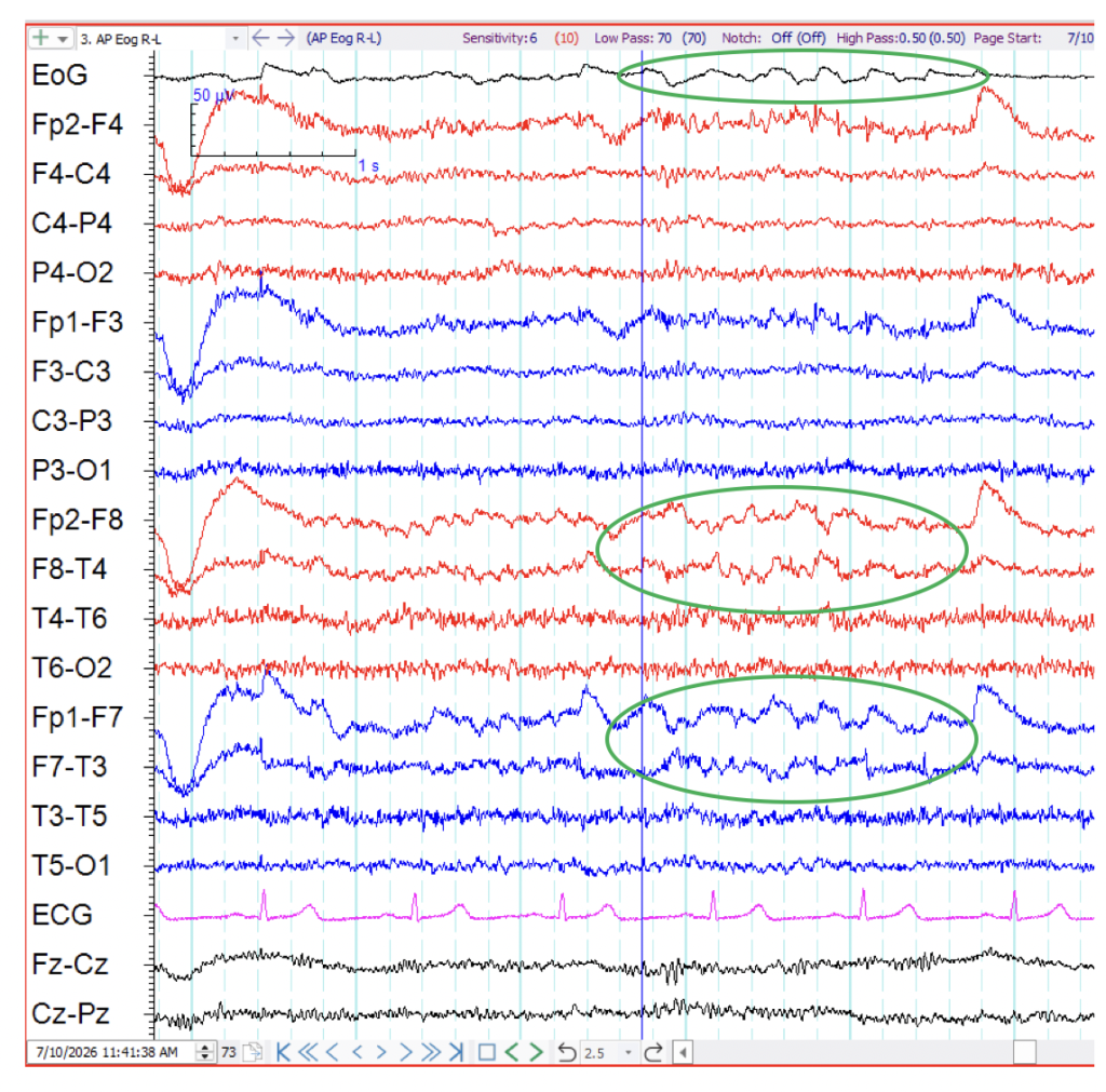
In the above EEG example the first channel is recording from the right outer canthus to the left outer canthus, showing eye movement activity. This helps differentiate eye movement from EEG activity on the EEG recording channels.
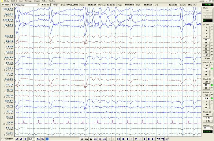
A more comprehensive study of eye movements can be done by adding EOG electrodes under each eye and creating a montage in relation to the electrodes above the eyes.
Below Right eye – A2
Fp2 – A2
Below Left eye – A1
Fp1 – A1
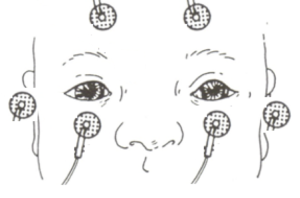
The following example shows phase reversing activity (out of phase) across the eyes synchronized with the frontal slow activity seen in the EEG. This suggest the frontal slowing activity is most likely caused from eye movement potential.
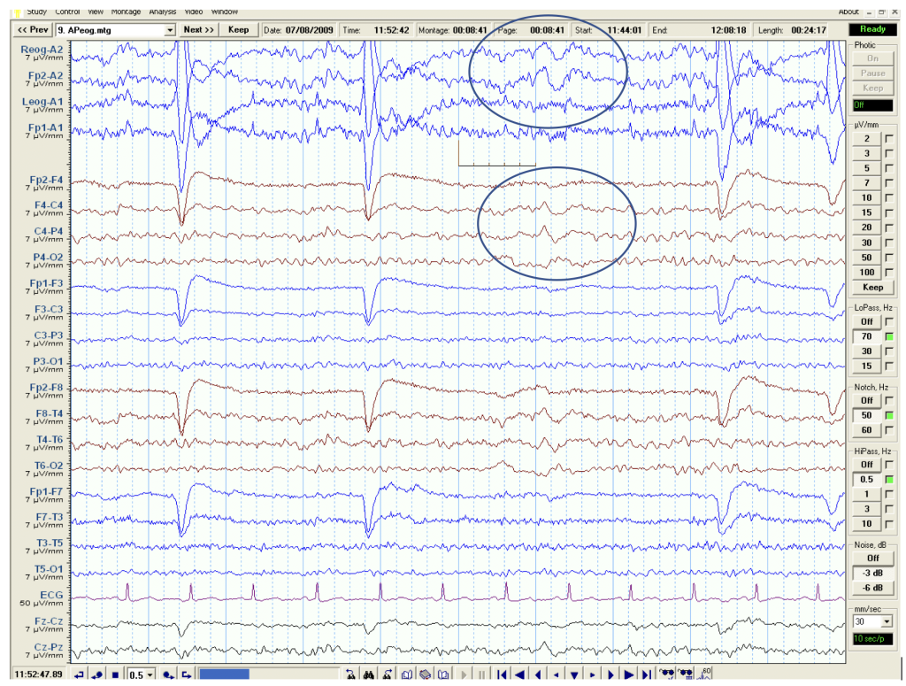
The next EEG example shows in phase activity recorded from the Right REOG electrode and from Fp2. This suggests the right frontal slow wave activity in the EEG is coming from the brain, not the eye potential.
EMG – Electromyograph
Additional EMG electrodes can be placed over any muscle. This can be useful to correlate specific movements such as myoclonic jerks to the EEG.
Add an example of EMG movement assisting with EEG interpretation.
More detail on the different physiological measurements and their recording parameters are discussed in the ANSA Learning short course Other Physiological Measurement Techniques (availability TBA).
