
“One man’s treasure is another man’s garbage”. In this topic activity that arises from the environment, the equipment or from the patient that is not wanted in the EEG recording is discussed. Artefacts can inhibit important waveform in the EEG.
From Merriam-Webster’s Medical Dictionary, 2002, the word ‘artefact’ has the following meanings:
- a product of artificial character due to extraneous (as human) agency; specifically : a product or formation in a microscopic preparation of a fixed tissue or cell that is caused by manipulation or reagents and is not indicative of actual structural relationships
- an electrocardiographic and electroencephalographic wave that arises from sources other than the heart or brain
In general, when recording an EEG, artefacts are signals that interfere with the EEG signals. In EEG recording, differential amplifiers are used to avoid as much interference as possible (see ANSA Learning short course Neurophysiology Amplifiers, Filters & Output). Not all artefacts present themselves equally to the amplifier so will not be eliminated with this method of amplification.
Recognising artefacts
Because the EEG signal is significantly amplified for the display system to produce an adequately sized deflection, it lends itself to amplifying other unwanted signals as well. Recognition of artefacts is an important part of the EEG recording. Many artefacts mimic EEG activity and can easily be mistaken as an abnormality.
The environment in which the EEG is recorded is often the source of interference. Except in the instance of recording in an open field, the electromagnetic field of mains frequency is always present. Other environment artefacts include electrostatic interference and radio frequency artefacts.
Instrumental artefacts arise from the machine itself or from the electrodes. The principles behind instrumental and electrode artefacts are discussed in the ANSA Learning short courses Neurophysiology Recording Electrodes, Neurophysiology Amplifiers, Filters & Output and EEG Electrode Application & Electrode Impedance. Electrode artefacts occur mainly due to damaged connections, inferior recording qualities or from poor application.
When recording an EEG, muscle activity (EMG), cardiac activity (ECG), sweat sway and other physiological measures are also considered artefacts.
An unusual waveform in the EEG should be considered artefact until proven otherwise.
Here are some examples of common physiological and environmental artefacts seen during an EEG.
Physiological artefacts:
Eye movement artefacts
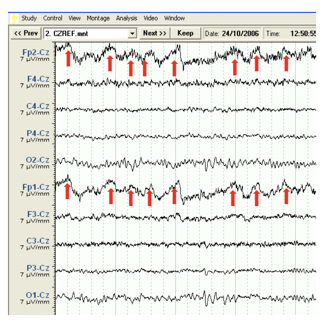
Blinking – Almost rhythmical 3Hz frontal activity caused by eye blinking can easily be mistaken for frontal EEG slowing to the untrained eye. In this example the artefact is seen only in the Fp2-Cz and Fp1-Cz channels so an assumption can be made that the potential is arising from the blinking movement of the eye.
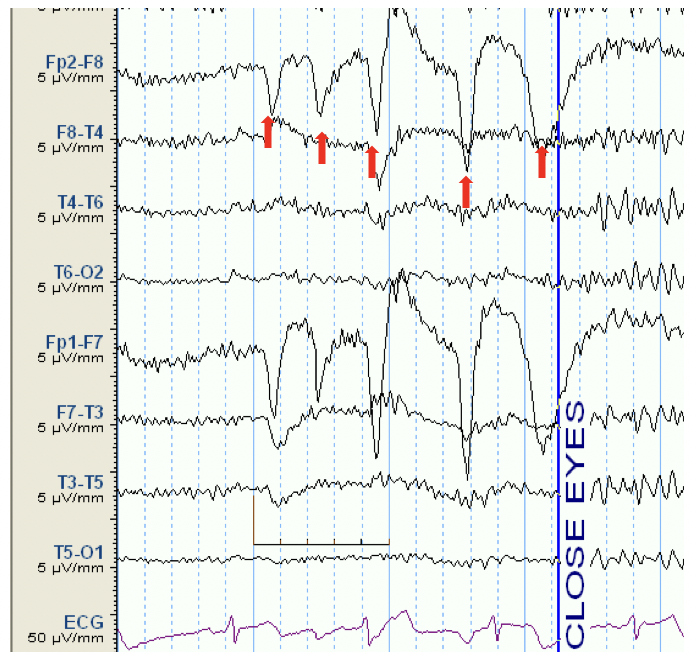
Blinking – Large blinks, especially when the eyes are open can mar the EEG recording. High amplitude eye blinks, such as these in the above example can mimic sharp waves.
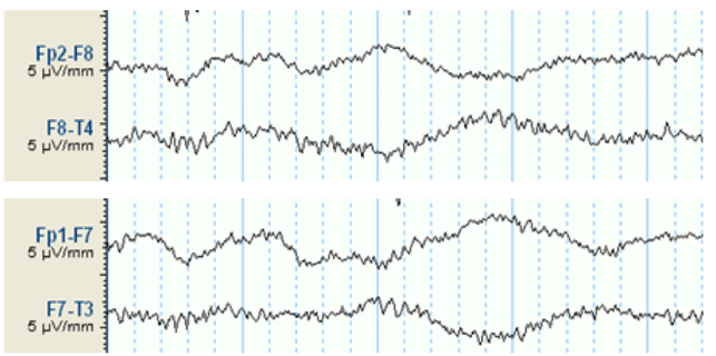
Rolling eye movements create the sway seen in the above example. As the eyes move from one side to the other the phase shifts from side to side on the EEG recording.
EMG related artefacts
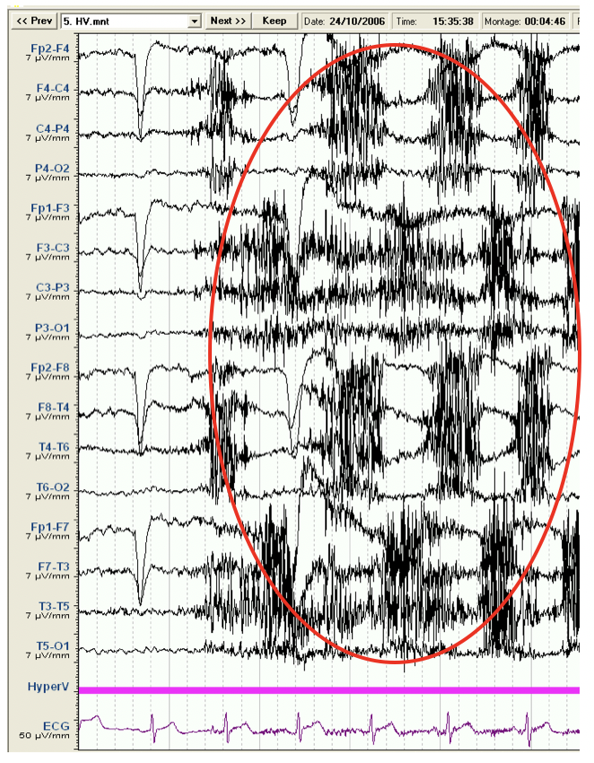
Clenching teeth/grinding movements
The dark high frequency, high amplitude EMG activity mars the underlying EEG. Clenching and grinding the teeth contracts the muscles on the scalp making it difficult to see the underlying EEG activity.
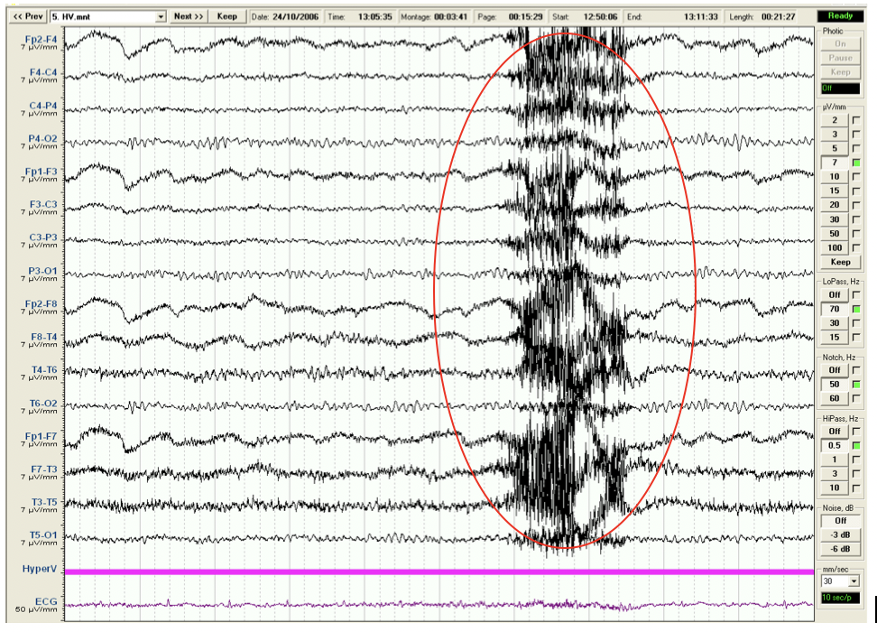
Swallow creates EMG activity that can be seen in a generalised distribution, significantly marring the background EEG.
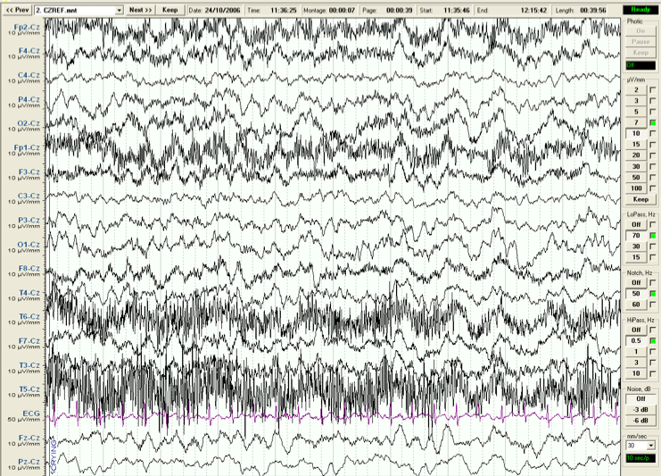
Crying also produces EMG artefact. In this example the EMG artefact is localised to the frontal and temporal regions seen prominently in the T6-Cz and T5-Cz channels and less in Fp2-Cz and Fp-Cz. It is significant enough to mar the background EEG in those regions.
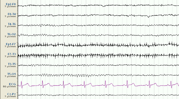
Small muscle fibre firing creates small fast potentials. In this example the muscle potential arises from F7.
Heart related artefacts
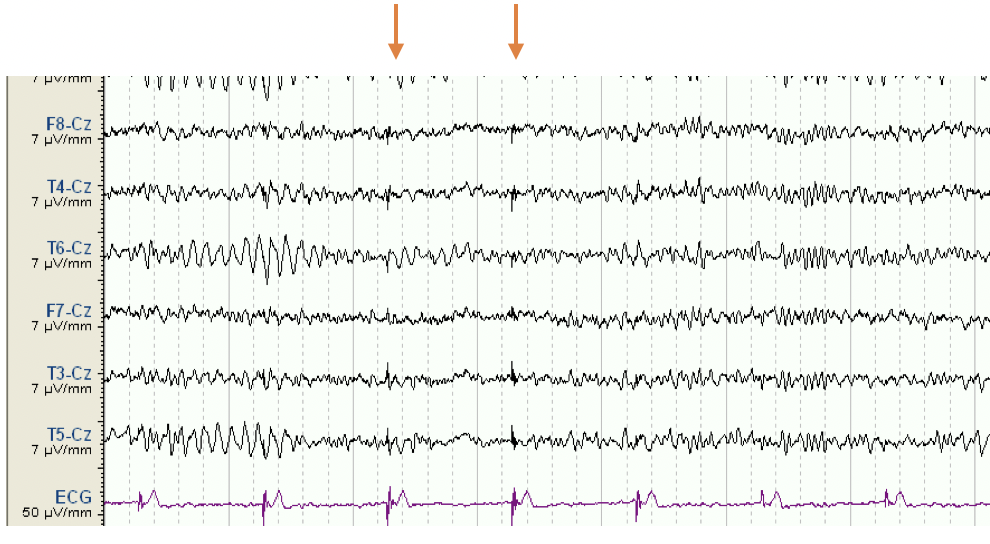
ECG Artefact
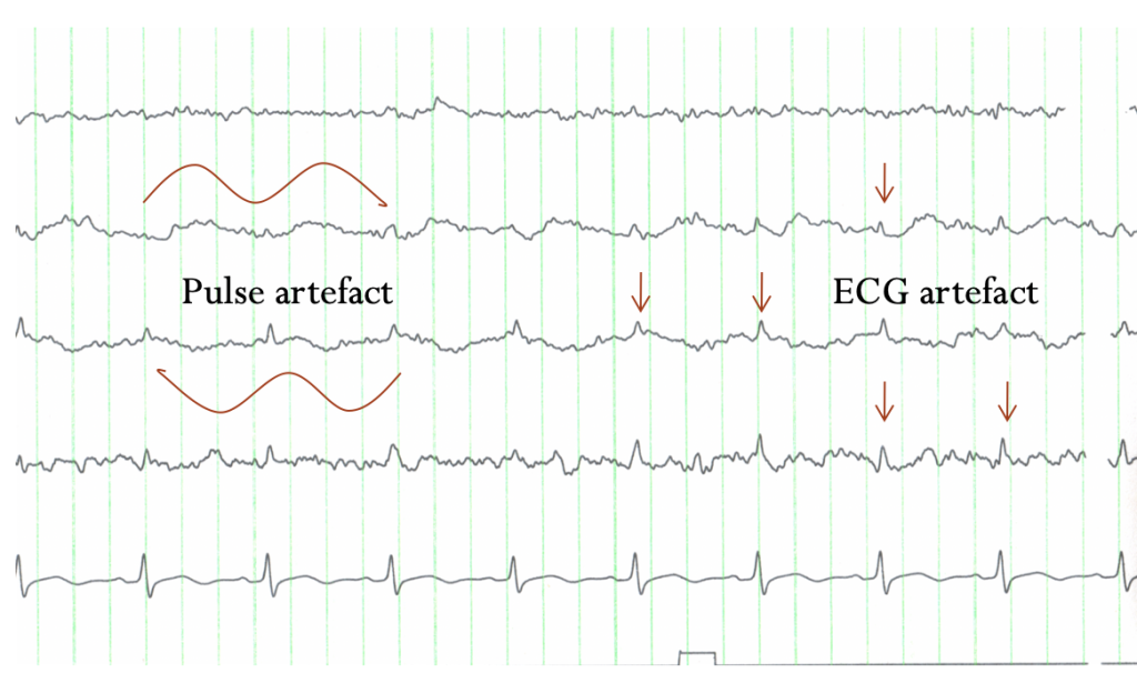
The ECG potential is a large potential with respect to EEG activity and can sometimes be seen in the EEG channels. Artefacts that are synchronised with the heart rate can be assumed to arise from the ECG or heart related phenomenon.
Pulse Artefact
Pulse artefact can be seen on the EEG recording when an electrode lies over a blood vessel.
In the example above pulse artefact can be seen in the 2nd and third channel as phase reversing slow wave activity that occurs at the same frequency of the heart rate.
This example also shows ECG artefact on almost all EEG channels.
Movement artefacts:
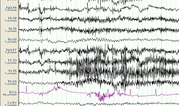
Movement of the patient causes EMG artefact seen in the left temporal region in this example. It and can also cause electrodes to become unstable, as seen here in the ECG lead.
Electrode artefact:
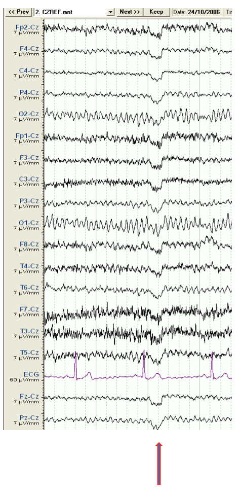
Contaminated reference
This example above shows the same unusual wave form on every channel. In this common reference montage it is likely this is an artefact arising from the Cz electrode.
Pop artefact
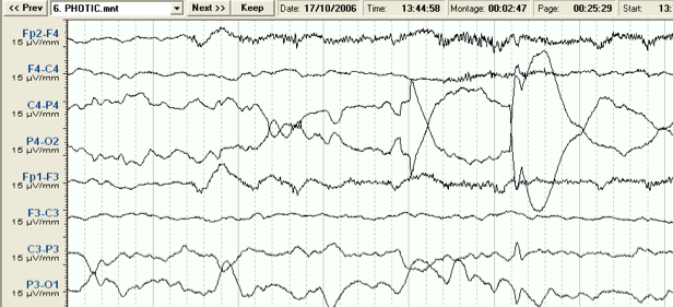
When the electrode is poorly applied and becomes unstable a sudden change in electrode contact can cause a difference in electrode potential, creating the artefact known as ‘Pop’ artefact.
Environmental artefact:
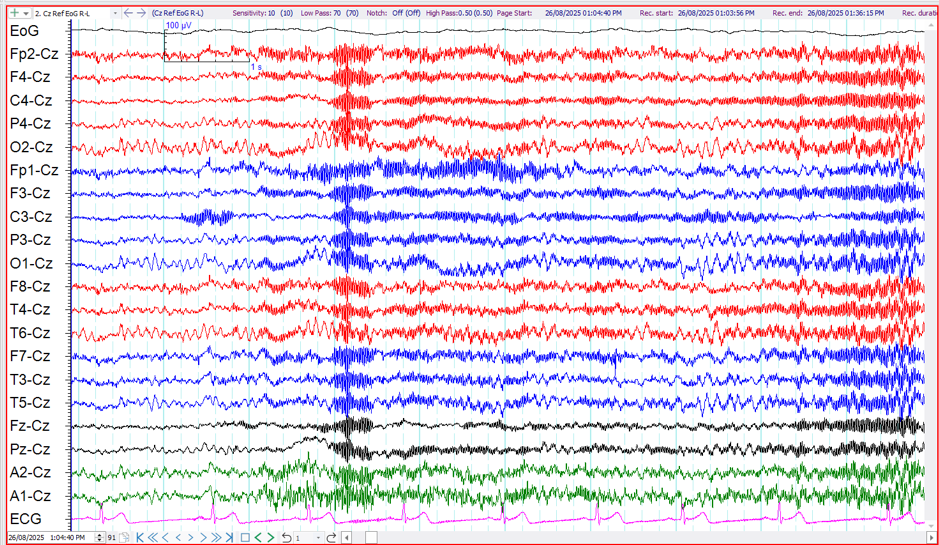
50Hz artefact
The most common environmental artefact seen on an EEG arises from the mains. Australian mains frequency is 50Hz.
Eliminating artefacts
It is important to reduce artefacts as much as possible without using the filters. Filters may attenuate, or reduce, certain artefacts but will also act in the same way on the EEG. There are many ways to reduce artefacts before the need to adjust the filters. The correct choice of electrodes and good application is essential. Remove nearby electrical equipment and turn off any unnecessary machinery. Patient relaxation techniques are also an essential part of removing artefacts.
Movement Artefacts
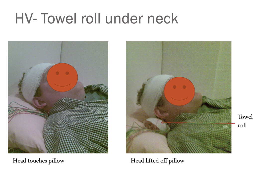
Sometimes just repositioning the patient slightly can eliminate the movement artefact. Raising the head off the pillow with extra support behind the neck or even asking the patient to turn their head to one side can reduce artefacts caused from the pillow contact. examples of simple artefact remedies are seen below.
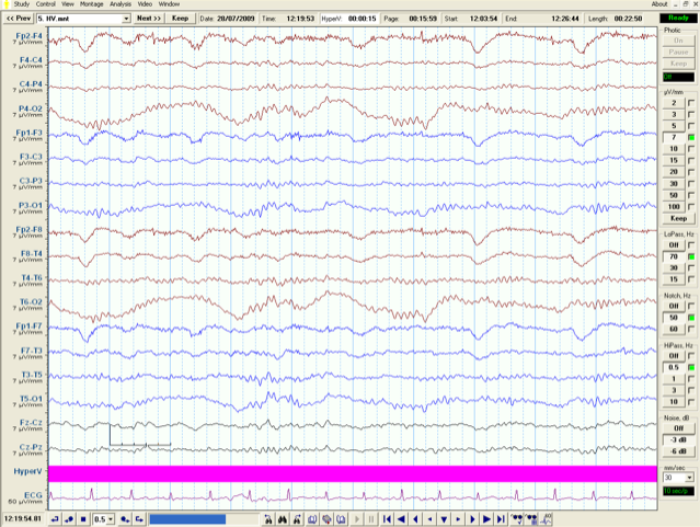
Head movement artefact during hyperventilation.
In the example above, a large slow potential is seen in the posterior head regions. This is a common movement artefact occurring as the patient moves their head with each deep breath during the hyperventilation exercise. This movement artefact can be reduced or eliminated simply by placing a rolled towel under the patient’s head. This stops the occipital electrodes moving on the pillow as the patient moves their head.
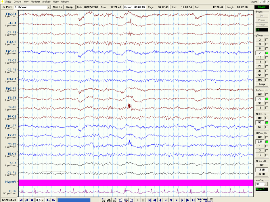
The posterior movement artefact has now stopped.
Electrode artefact
Electrodes may dislodge at any time if not secured well. A quick adjustment can eliminate artefact localised to one electrode.
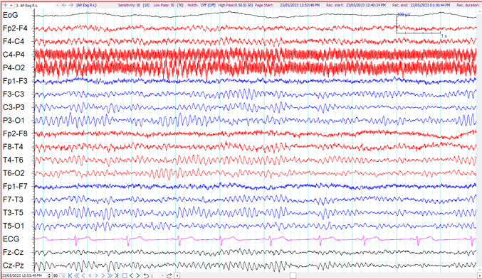
In the example above there is significant 50Hz artefact appearing in the C4-P4 and P4-O2 channels. We can assume the artefact is coming from P4, the common electrode to these two channels. Be re-securing the electrode the 50Hz artefact can be eliminated or reduced significantly.
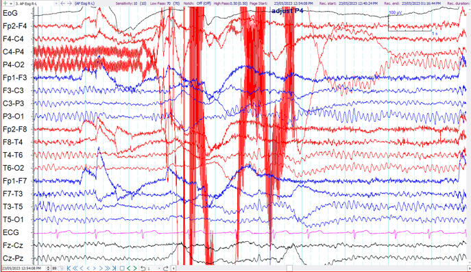
Once the electrode has been secured the 50Hz activity at P4 has reduced significantly.
Environmental artefact
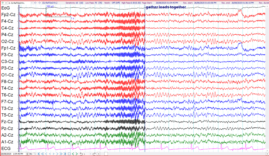
The above example shows 50Hz interference in all channels of the EEG recording. This can be caused by electromagnetic loops created by the EEG electrode leads. By gathering the electrodes together the 50Hz artefact has been eliminated.
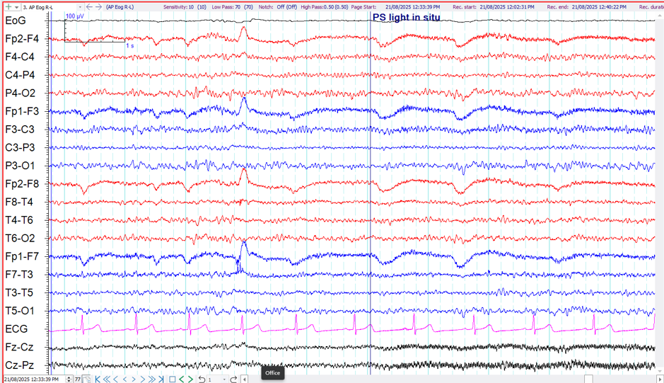
Other causes of 50Hz interference can be from electrical appliances in the nearby vicinity.
The example above shows the introduction of 50Hz interference when the Photic Strobe light is placed in front of the patient. This is particularly noticeable arising from Cz in this example.
Annotation of artefacts
For the experienced EEG recorder or reader, most artefacts are usually easy to spot. However there are many artefacts that mimic EEG activity or are difficult to recognise, particularly if you are not the one performing the EEG. For this reason it is important to make a note on the EEG when an artefact is recognized. Elimination of the artefact should also be noted. In this way the reader of the EEG can identify the true EEG waveforms from the artefact.
In the example above the source of the artefact is identified by making note of when the photic strobe light is placed in front of the patient. At that very same time 50Hz appears on the EEG recording.
Below are some more examples of recognising, eliminating and annotating artefacts.
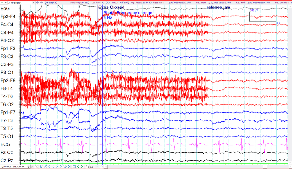
The example above shows EMG artefact on most EEG recording channels on the right side, more so in the right temporal region. When a patient has clenched teeth, the muscles in the temporal region are easily activated. By asking the patient to open their mouth slightly and not have their teeth touching, this type of artefact can often be eliminated, as is clearly annotated on this example.
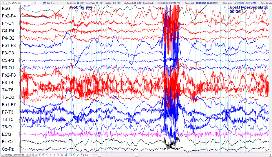
The above example shows an EEG sample with a number of artefacts including EMG activity and eye blinking. A symmetrical alpha rhythm can be seen in the occipital region as well as some rhythmical 3Hz slowing in the left posterior temporal/occipital region (T5-01). Is this rhythmical delta slowing? The annotation on the sample tells us the patient is rubbing their eye at this particular time. It can then be deduced that this rhythmical activity is mostly likely movement artefact.
